Recording Neuron Activity using Implantable Fluorescence Sensors
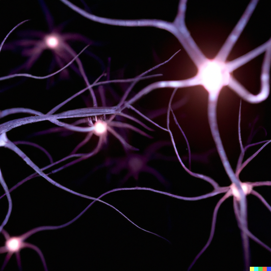
Neurostimulation is an effective way to modulate the nervous system to treat diseases and disorders like drug-resistant epilepsy, depression, and Parkinson’s. Stimulation acts by creating a local electric field with an electrode outside of an axon, inducing changes in voltage-gated ion channels that create an action potential (AP). For stimulation to be effective, the strength of the created electric field creates a Goldilocks problem – too small of an amplitude fails to activate the axon(s) in question and too large of an amplitude activates undesired axons that can signal the wrong direction or stimulate unintended organs and muscles. Electrical recordings or muscle twitches are used to find stimulation thresholds (by ramping up stimulation amplitude until AP is seen) but recording electrodes must be placed far from stimulation electrodes to avoid stimulation artifact, increasing the implant footprint (larger devices block more blood flow, creating necrotic tissue and eliciting more inflammatory response). We are working on a system that records action potentials chemically by measuring neurotransmitter and ion concentrations at synapses of interest. Using fluorophore-embedded waveguides and implantable miniaturized optics and electronics, we are working to improve the Bionode to both stimulate and record neurons simultaneously.